How Mole Check with Dermatoscope Done Is Painful?
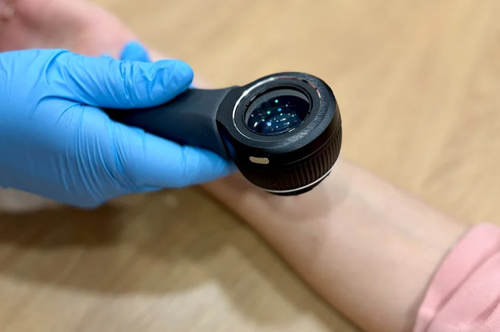
Photo: adoria.lv
Preventive mole check most effective way timely notice potentially dangerous changes as early detection main prerequisite successful treatment. Modern medicine for this purpose offers completely painless very precise method – dermatoscopy – providing immediate results preventing unnecessary anxiety.
About how this check done who needs it why definitely no need fear dermatoscopy continues to tell Health and Beauty Center Adoria dermatologist Jevgenija Čumakova.
In this article you'll learn:
- Dermatoscopy briefly: is this diagnostic method painful how works?
- Examination process: who needs prepare before visit?
- After check: what action after receiving check results?
What dermatoscopy how helps diagnose skin formations?
Dermatoscopy non-invasive (performed without surgical intervention) completely painless quick skin formations diagnostic method allowing obtain much more detailed information than possible see with naked eye. During used special instrument – dermatoscope – equipped with magnifying glass polarized light source.
This technology allows dermatologist view not only skin's upper layer (epidermis) but also deeper skin layers – dermis. Light emitted through dermatoscope illuminates skin structures pigmentation blood vessel network other signs not visible in usual examination. Thus possible much more precisely evaluate moles symmetry borders color variations structure distinguishing benign formations from potentially malignant.
Dermatoscopy basis for early melanoma other skin tumors diagnostics. If patient timely turns to doctor with this examination help possible notice suspicious changes at early stage. At this stage treatment ensures most effective results significantly reducing both complication risk negative impact on patient's quality of life.
- How distinguish which moles potentially dangerous how perform skin formations self-control at home using ABCDE method recommended performed by dermatologists worldwide? Learn all reading article "Moles Skin Formations: When Need Check by Dermatologist?".
How often recommended perform mole check?

Photo: freepik.com/Freepik
Preventive mole check recommended perform at least once a year everyone even if no complaints visible changes. However exist certain risk groups whom checks should be done more often – every six months or according to dermatologist individually created control check schedule.
Elevated risk factors include:
- Fair skin phototype (I or II): people with fair skin blue green eyes blond red hair have higher sunburn skin cancer risk.
- Large number of moles (usually over 50): clinically proven large total number of moles increases skin tumor development risk. Doesn't mean each individual mole dangerous but overall skin condition monitoring needs enhanced attention.
- Atypical dysplastic moles appearance: recognizable by irregular borders multiple color shades asymmetric shape higher risk become malignant. Itself such formation not melanoma but indicates need particularly thorough regular monitoring.
- Skin cancer in family history: if patient herself first-degree relatives (parents siblings) had melanoma other skin tumor.
- Frequent intense sunburns: especially in childhood teenage years obtained sunburns with skin peeling.
- Regular solarium visits or prolonged sun exposure due to work specifics.
Why dermatologist best specialist for mole assessment?
Though regular self-examination important full adequate precise mole evaluation can perform only qualified dermatologist. Skin formations naked eye may seem similar in dermatologist's view reveal very different signs. Thanks to experience specialized technologies e.g., dermatoscope specialist able notice finest structure changes indicating early risk prevent unfounded anxiety about benign formations.
E.g., some dark skin formation can cause patient great concern but doctor with dermatoscope help able unmistakably identify as completely harmless seborrheic wart hemangioma pigment spot. Exactly this ability precisely distinguish different formations allows avoid unnecessary surgical procedures develop each case matching correct action plan.
- Sometimes hard understand whether with skin problem – pimples redness pigmentation spots – go to cosmetologist or still need dermatologist help. More about both specialists' differences how choose right read article "Dermatologist vs Cosmetologist: How Know Which Specialist Turn To?".
How mole check procedure itself done?
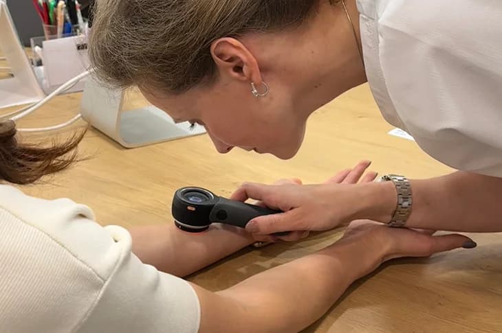
Photo: adoria.lv
Patient preparation for specialist visit minimal – preferable arrive with clean skin not using body lotions creams on examination areas as can create glare complicate examination. Procedure itself simple quick comfortable. Examination course can divided into several steps:
- Patient medical history collection: conversation with patient about skin condition previous diseases family anamnesis any noticed changes specific moles (e.g., growth color change itching bleeding).
- Visual examination: whole body skin examination gain overall picture of moles number location type. Also checked harder accessible places e.g., head hairy part feet skin between toes.
- Examination with dermatoscope: each formation causing even slightest doubt detailed studied with dermatoscope. Specialist applies to skin small amount gel oil improve optical visibility then attaches instrument to skin. This process completely painless causes no discomfort.
- Recommendations provision: after examination specialist explains results provides recommendations for further monitoring course or if needed orders additional examinations.
Is mole check needed also for children?
Though malignant skin tumors in children rare mole check can be necessary also in this age group. Children teenagers moles can appear new grow larger slightly change color under body growth influence usually normal process.
However if some formation grows disproportionately fast has irregular shape multiple colors starts cause discomfort definitely needed consultation. Dermatologist with experience working with children able professionally assess situation distinguish physiological growth signs from pathological calm parents. Regular checks in childhood also form correct health habits for future.
What expect after dermatoscopy examination?
Dermatoscopy examination ends with doctor's conclusion clear action plan. Based on examination results dermatologist detailed explains each suspicious formation condition provides individual recommendations for further action.
- All formations benign: if during examination no suspicious signs discovered recommended continue regular self-examination come for preventive check after year or according to individual risk profile.
- Found atypical but not malignant formation: sometimes mole has some atypical signs but insufficient diagnose malignant process. In such case dermatologist can recommend monitor formation creating its digital photo (so-called skin formation "passport") use as reference point for next checks ordering repeated visit after 3–6 months.
- Suspicion of malignant formation: if during check discovered malignant process characteristic signs dermatologist will recommend perform biopsy. Procedure during which suspicious formation surgically removed sent for histological examination to laboratory. This analysis where tissue sample studied at microscopic level only method allowing one hundred percent confirm deny tumor diagnosis.
Don't Risk Book Preventive Mole Check!

Photo: adoria.lv
Don't postpone skin health care for later – timely professional check safest way take care of your health gain peace of mind. Experienced dermatologists modern diagnostic methods dermatoscopy individual approach to each patient – all offered at Health and Beauty Center Adoria. Book your visit adoria.lv or call +371 67 315 000!