Dopplerography During Pregnancy: Why Important?
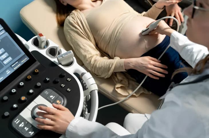
Photo: freepik.com/Freepik
Pregnancy special time in every woman's life when concerns for expected baby's health become priority. Modern medicine provides opportunity carefully monitor fetal development prospective mother's health offering various diagnostic methods. Though ultrasound for pregnant women indispensable method for fetal development assessment dopplerography offers in-depth insight exactly into blood flow indicators providing essential additional information about expected baby's health condition.
Why dopplerography examinations have crucial role in pregnancy monitoring what data they provide – continues to tell Health and Beauty Center Adoria gynecologist Natālija Gailīte.
Most Important About Dopplerography During Pregnancy:
- Blood Flow Assessment: dopplerography provides detailed fetal placenta uterine blood flow analysis.
- Risk Diagnostics: examination allows timely detect possible pregnancy complications.
- Care Optimization: provides essential information for further pregnant women care plan development.
What Dopplerography How Works?
Dopplerography non-invasive ultrasound examination method allowing in real time assess blood flow speed direction in various blood vessels. Based on Doppler effect – sound wave frequency changes when reflected from moving objects in this case – from red blood cells (erythrocytes).
- While standard ultrasonography primarily reveals information about organ structure anatomical features dopplerography's main task assess blood flow functional state.
During procedure specialist uses ultrasound probe directing high-frequency sound waves through body tissues. Upon contact with blood cells flowing in blood vessels these waves reflected back to probe. Computer analyzes reflected wave frequency changes converts them into color image graphical curve depicting blood flow speed direction resistance in specific blood vessels.
Performing examination in pregnant women most often studied blood flow in umbilical cord arteries vein fetal middle cerebral artery fetal ductus venosus pregnant woman's uterine arteries. This information extremely valuable assess placenta function fetal supply with oxygen nutrients identify possible risks to fetal development.
Dopplerography examination safe for both pregnant woman expected baby as no ionizing radiation used. Procedure usually lasts from 15 to 30 minutes no special preparation needed before examination.
- How ultrasound examinations can help identify organ structural changes potentially indicating possible hormonal disorders? Learn reading article: "Does Ultrasonography Help Detect Hormonal Changes?".
Dopplerography Importance in Different Pregnancy Stages

Photo: adoria.lv
Unlike standard examinations intended for all pregnant women dopplerography targeted diagnostic method used in certain situations pregnancy stages obtain additional information about fetal health placenta functions.
First Pregnancy Trimester
In first pregnancy trimester dopplerography usually not performed unless specific medical indications e.g., suspicion of ectopic pregnancy other situations but during first trimester ultrasound screening possible perform uterine artery doppler examinations allowing assess preeclampsia risk.
Second Pregnancy Trimester
In second trimester usually between 20th 24th pregnancy week can be performed uterine artery dopplerography. This examination helps assess blood flow in arteries supplying uterus placenta. Elevated resistance in these blood vessels can indicate increased risk develop preeclampsia fetal growth restriction in later pregnancy stages. Such information allows timely plan adjusted pregnant care initiate preventive measures if necessary.
Third Pregnancy Trimester
Most often with greatest importance dopplerography used in third pregnancy trimester especially if suspicion of fetal growth restriction reduced amniotic fluid amount if mother has some chronic diseases e.g., diabetes arterial hypertension in multiple pregnancy if previous pregnancies had complications. In this period dopplerography helps assess:
- umbilical cord artery blood flow: provides information about placenta's ability deliver fetus oxygen nutrients. Pathological blood flow can indicate severe placental insufficiency fetal endangerment.
- fetal middle cerebral artery blood flow: helps diagnose fetal hypoxia (oxygen deficiency) anemia.
- ductus venosus blood flow: allows assess fetal heart function venous return potentially disturbed in pronounced hypoxia.
Timely obtained dopplerography data allows specialists make informed decisions about further pregnancy care plan need for additional examinations hospitalization even preterm labor induction ensure best possible outcome for both prospective mother expected baby.
What Risks Dopplerography Helps Detect?
Modern pregnancy care based on ability proactively identify manage possible risks ensuring best possible outcome. Dopplerography in this regard invaluable additional diagnostic method timely allowing detect various potential threats to normal fetal development health.
- Fetal growth restriction: one of main conditions dopplerography helps diagnose fetal growth restriction (IUGR). If placenta unable provide sufficient nutrient oxygen supply fetal growth slows. Dopplerography especially umbilical cord artery examination can show elevated blood flow resistance even reverse (opposite) blood flow in diastole phase indicating severe placental failure fetal endangerment.
- Preeclampsia risk: as already mentioned uterine artery dopplerography in first second trimester helps identify pregnant women with elevated preeclampsia risk. Timely risk group determination allows initiate preventive measures e.g., low-dose aspirin use potentially reducing preeclampsia development likelihood severity.
- Placental insufficiency: dopplerography indispensable method also for placental insufficiency diagnostics. This condition can cause not only fetal growth restriction but also chronic hypoxia negatively affecting fetal organ development potentially leading to serious health problems after birth.
- Fetal anemia: examining fetal middle cerebral artery possible diagnose fetal anemia. In anemia case blood flow speed in this artery increases. Important e.g., in Rh conflict other hematological problems.
- Multiple pregnancy: such pregnancies associated with increased specific complication risk dopplerography critically important timely diagnose conditions like twin-twin transfusion syndrome (TTTS) selective fetal growth restriction (sIUGR) twin anemia-polycythemia sequence (TAPS). Allows individually assess each fetus's blood flow condition well-being optimizing care.
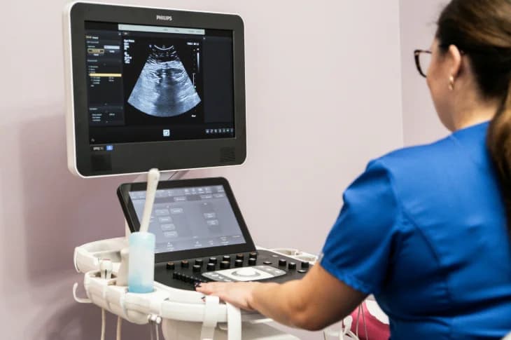
Photo: adoria.lv
Overall dopplerography provided information helps individually tailor pregnancy monitoring optimize treatment plan timely respond to possible complications thus significantly improving pregnancy course.
- What role of gynecological ultrasonography in reproductive health care prevention read our specialists prepared article: "What Gynecological Ultrasonography When Needed?".
Modern Gynecology in Riga Health and Beauty Center Adoria
Gynecologist consultations modern reproductive health pregnant women care gynecological pregnant women ultrasound with latest generation diagnostic equipment experienced specialists Health and Beauty Center Adoria gynecology in Riga A. Čaka street 70-3. Book your visit now!